
Fly microscopy study outline
Section A: Fly anatomy under magnification
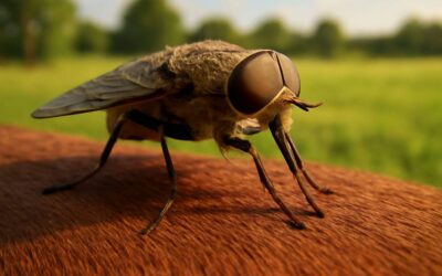
Tiny world, big details—flies under microscope reveal a world where every bristle and pore has a job. Section A surveys anatomy through a lens that makes miniature architecture feel dramatically cinematic. I love how this lens makes the clockwork of a millimeter feel like a heist movie cameo.
Under magnification, the head hosts compound eyes, antennae, and a streamlined proboscis. The thorax anchors two wings and three pairs of legs; halteres act as tiny gyroscopes. The abdomen houses organ systems and a payload of sensory data.
- Head features: compound eyes, antennal bases, mouthparts
- Thorax and wings: wing venation, halteres, leg articulation
- Abdomen and sensory surfaces: segmentation, bristles, pores
Observing with patience, you notice the micro sculpting of the exoskeleton and how light plays across the cuticle. Here in South Africa’s labs, every glide of a bristle is a note in the instrument of flight!
Section B: Microscopy techniques for studying flies
One photon can unveil a thousand stories, and Section B translates that whisper into technique. In the quiet hum of South Africa’s labs, flies under microscope become a poetry of light and shadow, where contrast and depth sculpt the narrative of tinier anatomy without losing the atmosphere.
Technique is the instrument here—lighting schemes and optical modes that tease texture from the exoskeleton and surface detail without breaking the mood.
- Brightfield and darkfield illuminate general morphology with shadow and glow.
- Differential interference contrast teases microtopography from the cuticle, like a whisper in stone.
- Fluorescence and confocal methods map labeled tissues while preserving the drama under the lens.
These approaches let the observer hear the minute clockwork without shouting, a balance that suits South Africa’s meticulous, moonlit laboratories.
Section C: Research applications of microscopy in flies
Section C anchors the study of flies in a practical horizon, where microscopy translates promise into practice. The fruit fly brain holds about 100,000 neurons—a compact cosmos hinting at universal principles. In the quiet glow of South African laboratories, flies under microscope become a storyboard of development, signaling, and behavior, where subtle contrast teases timing and fate without shouting.
Key research avenues unfold with clarity and care:
- Neural circuitry mapping and functional assays
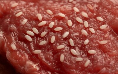
- Developmental timing and morphogenesis
- Toxicology and pharmacology screening
- Disease modelling and behavioral phenotyping
From these applications, science threads a bridge between bench and broader understanding. Flies under microscope reveal how genes sculpt form, regulate timing, and expose vulnerability to environmental cues. The cadence remains precise, dramatic, and practical, inviting collaboration across disciplines while honoring the tiny life that casts long shadows across the lab bench.
Section D: Educational and DIY microscopy projects with flies
A single fruit fly under a disciplined lens becomes a compass for wonder and inquiry. In classrooms across South Africa, ‘Tiny teachers, huge lessons’ guide students as they observe growth, timing, and behavior with quiet precision. flies under microscope invites hands-on learning that feels magical yet rigorously grounded, turning abstract ideas into living stories you can almost hear.
Educators curate modules that are not about expensive gear but about storytelling through light and time.
- Accessible, low-cost optics and smartphone adapters for little labs
- Concept-driven explorations of life stages, neurons, and behavior
- Citizen science collaborations linking schools with local research centers
These gentle, DIY investigations invite learners to contribute their own observations, weaving patience and wonder into every slide and shadow.
Section E: Comparative entomology: flies vs other insects under magnification
Comparative entomology under magnification is where elegance meets elbow room for curiosity! When you glimpse flies under microscope, their tiny differences become grand conversations about life histories, wing choreography, and sensory tricks that invite sharper questions than a lecture hall sigh.
Under magnification, wing venation, antennae style, and mouthparts reveal contrasts that matter to ecology and behavior. Flies, with trimmed, feathery antennae, behave differently from beetles with hard elytra and wasps with slender waists; the frames tell stories before a single word is spoken.
- Wing architecture and venation patterns
- Antennae form and sensory layout
- Mouthparts and feeding strategies
These insights enrich classroom dialogue, making science social, not siloed, and perfectly suited to South Africa’s diverse ecosystems.


0 Comments